Monday, January 30, 2023

Proteomic discovery of chemical probes that perturb protein complexes in human cells

Michael R. Lazear, Jarrett R. Remsberg, Martin G. Jaeger, Katherine Rothamel, Hsuan-lin Her, Kristen E. DeMeester, Evert Njomen, Simon J. Hogg, Jahan Rahman, Landon R. Whitby, Sang Joon Won, Michael A. Schafroth, Daisuke Ogasawara, Minoru Yokoyama, Garrett L. Lindsey, Haoxin Li, Jason Germain, Sabrina Barbas, Joan Vaughan, Thomas W. Hanigan, Vincent F. Vartabedian, Christopher J. Reinhardt, Melissa M. Dix, Seong Joo Koo, Inha Heo, John R. Teijaro, Gabriel M. Simon, Brahma Ghosh, Omar Abdel-Wahab, Kay Ahn, Alan Saghatelian, Bruno Melillo, Stuart L. Schreiber, Gene W. Yeo, Benjamin F. Cravatt

bioRxiv 2022.12.12.520090; doi: https://doi.org/10.1101/2022.12.12.520090

Molecular Cell, 2023

https://doi.org/10.1016/j.molcel.2023.03.026

Most human proteins lack chemical probes, and several large-scale and generalizable small-molecule binding assays have been introduced to address this problem. How compounds discovered in such “binding-first” assays affect protein function, nonetheless, often remains unclear. Here, we describe a “function-first” proteomic strategy that uses size exclusion chromatography (SEC) to assess the global impact of electrophilic compounds on protein complexes in human cells. Integrating the SEC data with cysteine-directed activity-based protein profiling identifies changes in protein-protein interactions that are caused by site-specific liganding events, including the stereoselective engagement of cysteines in PSME1 and SF3B1 that disrupt the PA28 proteasome regulatory complex and stabilize a dynamic state of the spliceosome, respectively. Our findings thus show how multidimensional proteomic analysis of focused libraries of electrophilic compounds can expedite the discovery of chemical probes with site-specific functional effects on protein complexes in human cells.


Monday, January 23, 2023

Proximity-labeling chemoproteomics defines the subcellular cysteinome and inflammation-responsive mitochondrial redoxome

Tianyang Yan, Ashley R Julio, Miranda Villanueva, Anthony E Jones, Andréa B Ball, Lisa M Boatner, Alexandra C Turmon, Stephanie L Yen, Heta S Desai, Ajit S Divakaruni, Keriann M Backus

bioRxiv 2023.01.22.525042; doi: https://doi.org/10.1101/2023.01.22.525042

Proteinaceous cysteines function as essential sensors of cellular redox state. Consequently, defining the cysteine redoxome is a key challenge for functional proteomic studies. While proteome-wide inventories of cysteine oxidation state are readily achieved using established, widely adopted proteomic methods such as OxiCat, Biotin Switch, and SP3-Rox, they typically assay bulk proteomes and therefore fail to capture protein localization-dependent oxidative modifications. To obviate requirements for laborious biochemical fractionation, here, we develop and apply an unprecedented two step cysteine capture method to establish the Local Cysteine Capture (Cys-LoC), and Local Cysteine Oxidation (Cys-LOx) methods, which together yield compartment-specific cysteine capture and quantitation of cysteine oxidation state. Benchmarking of the Cys-LoC method across a panel of subcellular compartments revealed more than 3,500 cysteines not previously captured by whole cell proteomic analysis, together with unexpected non-organelle specific TurboID-catalyzed proximity labeling. This mislabeling was minimized through simultaneous depletion of both endogenous biotin and newly translated TurboID fusion protein. Application of the Cys-LOx method to LPS stimulated murine immortalized bone marrow-derived macrophages (iBMDM), revealed previously unidentified mitochondria-specific inflammation-induced cysteine oxidative modifications including those associated with oxidative phosphorylation. These findings shed light on post-translational mechanisms regulating mitochondrial function during the cellular innate immune response.


Sunday, January 22, 2023

Covalent chemical probes for protein kinases

Ricardo A.M. Serafim, Lisa Haarer, Júlia G.B. Pedreira, Matthias Gehringer,

Current Research in Chemical Biology, 2023, 3, 100040,

https://doi.org/10.1016/j.crchbi.2022.100040

Small-molecule chemical probes are crucial tools to study the function of unexplored proteins in biological systems, thereby directly impacting preclinical target validation. Being one of the largest protein families in humans, protein kinases are currently among the most important and fruitful molecular targets in drug discovery. However, a significant fraction of the human “kinome” is still understudied and growing efforts in the scientific community aim at the development of specific chemical tool compounds for such “dark” kinases. Covalent targeting has proven to be a valid and rational strategy towards high-quality chemical probes enabling superior potencies, high selectivities and sustained target engagement. In the kinase field, the targeting of non-catalytic cysteine residues has been particularly fruitful and there is an increasing interest in addressing other residues, such as lysine or tyrosine. Herein, we discuss the properties and generation of covalent kinase inhibitors, with a special emphasis on electrophilic functional groups that can be used as “warheads”. Moreover, we highlight studies showcasing the development of covalent chemical probes targeting cysteine and lysine residues in an irreversible or reversible-covalent manner.


Saturday, January 21, 2023

Profiling Sulfur(VI) Fluorides as Reactive Functionalities for Chemical Biology Tools and Expansion of the Ligandable Proteome

 Katharine E. Gilbert, Aini Vuorinen, Arron Aatkar, Peter Pogány, Jonathan Pettinger, Emma K. Grant, Joanna M. Kirkpatrick, Katrin Rittinger, David House, Glenn A. Burley, and Jacob T. Bush

ACS Chemical Biology 2023
DOI: 10.1021/acschembio.2c00633

Here, we report a comprehensive profiling of sulfur(VI) fluorides (SVI-Fs) as reactive groups for chemical biology applications. SVI-Fs are reactive functionalities that modify lysine, tyrosine, histidine, and serine sidechains. A panel of SVI-Fs were studied with respect to hydrolytic stability and reactivity with nucleophilic amino acid sidechains. The use of SVI-Fs to covalently modify carbonic anhydrase II (CAII) and a range of kinases was then investigated. Finally, the SVI-F panel was used in live cell chemoproteomic workflows, identifying novel protein targets based on the type of SVI-F used. This work highlights how SVI-F reactivity can be used as a tool to expand the liganded proteome.


Friday, January 20, 2023

Fast and Bioorthogonal Release of Isocyanates in Living Cells from Iminosydnones and Cycloalkynes

Maxime Ribéraud, Karine Porte, Arnaud Chevalier, Léa Madegard, Aurélie Rachet, Agnès Delaunay-Moisan, Florian Vinchon, Pierre Thuéry, Giovanni Chiappetta, Pier Alexandre Champagne, Grégory Pieters, Davide Audisio, and Frédéric Taran

Journal of the American Chemical Society 2023
DOI: 10.1021/jacs.2c09865

Bioorthogonal click-and-release reactions are powerful tools for chemical biology, allowing, for example, the selective release of drugs in biological media, including inside animals. Here, we developed two new families of iminosydnone mesoionic reactants that allow a bioorthogonal release of electrophilic species under physiological conditions. Their synthesis and reactivities as dipoles in cycloaddition reactions with strained alkynes have been studied in detail. Whereas the impact of the pH on the reaction kinetics was demonstrated experimentally, theoretical calculations suggest that the newly designed dipoles display reduced resonance stabilization energies compared to previously described iminosydnones, explaining their higher reactivity. These mesoionic compounds react smoothly with cycloalkynes under physiological, copper-free reaction conditions to form a click pyrazole product together with a released alkyl- or aryl-isocyanate. With rate constants up to 1000 M–1 s–1, this click-and-release reaction is among the fastest described to date and represents the first bioorthogonal process allowing the release of isocyanate electrophiles inside living cells, offering interesting perspectives in chemical biology.


Monday, January 9, 2023

Discovery of Potent Small-Molecule Inhibitors of WDR5-MYC Interaction

Jian Ding, Guo Li, Hejun Liu, Lulu Liu, Ying Lin, Jingyan Gao, Guoqiang Zhou, Lingling Shen, Mengxi Zhao, Yanyan Yu, Weihui Guo, Ulrich Hommel, Johannes Ottl, Jutta Blank, Nicola Aubin, Yi Wei, Hu He, David R. Sage, Peter W. Atadja, En Li, Rishi K. Jain, John A. Tallarico, Stephen M. Canham, Ying-Ling Chiang, and He Wang

ACS Chemical Biology 2023

DOI: 10.1021/acschembio.2c00843

WD repeat domain 5 (WDR5) is a member of the WD40-repeat protein family that plays a critical role in multiple processes. It is also a prominent target for pharmacological inhibition in diseases such as cancer, aging, and neurodegenerative disorders. Interactions between WDR5 and various partners are essential for sustaining its function. Most drug discovery efforts center on the WIN (WDR5 interaction motif) site of WDR5 that is responsible for the recruitment of WDR5 to chromatin. Here, we describe the discovery of novel WDR5 inhibitors for the other WBM (WDR5 binding motif) pocket on this scaffold protein, to disrupt WDR5 interaction with its binding partner MYC by high-throughput biochemical screening, subsequent molecule optimization, and biological assessment. These new WDR5 inhibitors provide useful probes for future investigations of WDR5 and an avenue for targeting WDR5 as a therapeutic strategy.


Friday, January 6, 2023

Inhibiting androgen receptor splice variants with cysteine-selective irreversible covalent inhibitors to treat prostate cancer

Thirumagal Thiyagarajan, Suriyan Ponnusamy, Dong-Jin Hwang, Yali He, Sarah Asemota, Kirsten L Young, Daniel L Johnson, Vera Bocharova, Weidong Zhou, Abhinav K Jain, Emanuel F Petricoin 5, Zheng Yin, Lawrence M Pfeffer, Duane D Miller, Ramesh Narayanan 

PNAS, 120 (1) e2211832120

https://doi.org/10.1073/pnas.2211832120

Androgen receptor (AR) and its splice variants (AR-SVs) promote prostate cancer (PCa) growth by orchestrating transcriptional reprogramming. Mechanisms by which the low complexity and intrinsically disordered primary transactivation domain (AF-1) of AR and AR-SVs regulate transcriptional programming in PCa remains poorly defined. Using omics, live and fixed fluorescent microscopy of cells, and purified AF-1 and AR-V7 recombinant proteins we show here that AF-1 and the AR-V7 splice variant form molecular condensates by liquid–liquid phase separation (LLPS) that exhibit disorder characteristics such as rapid intracellular mobility, coactivator interaction, and euchromatin induction. The LLPS and other disorder characteristics were reversed by a class of small-molecule-selective AR-irreversible covalent antagonists (SARICA) represented herein by UT-143 that covalently and selectively bind to C406 and C327 in the AF-1 region. Interfering with LLPS formation with UT-143 or mutagenesis resulted in chromatin condensation and dissociation of AR-V7 interactome, all culminating in a transcriptionally incompetent complex. Biochemical studies suggest that C327 and C406 in the AF-1 region are critical for condensate formation, AR-V7 function, and UT-143’s irreversible AR inhibition. Therapeutically, UT-143 possesses drug-like pharmacokinetics and metabolism properties and inhibits PCa cell proliferation and tumor growth. Our work provides critical information suggesting that clinically important AR-V7 forms transcriptionally competent molecular condensates and covalently engaging C327 and C406 in AF-1, dissolves the condensates, and inhibits its function. The work also identifies a library of AF-1-binding AR and AR-SV-selective covalent inhibitors for the treatment of PCa.


Novel Irreversible Peptidic Inhibitors of Transglutaminase 2

Nicholas J. Cundy,   Jane Arciszewski,   Eric W. J. Gates,   Sydney L. Acton,   Kyle D. Passley,   Ernest Awoonor-Williams,   Elizabeth K. Boyd,   Nancy Xu,   Elise Pierson,   Catalina Fernandez-Ansieta,   Marie R. Albert,   Nicole M. R. McNeil,   Gautam Adhikary,   Richard L. Eckert  and  Jeffrey W Keillor  

RSC Medicinal Chemistry, 2023

DOI https://doi.org/10.1039/D2MD00417H

Transglutaminase 2 (TG2), also referred to as tissue transglutaminase, is an enzyme that plays crucial roles in both protein crosslinking and cell signalling. It is capable of both catalysing transamidation and acting as a G-protein, these activities being conformation-dependent, mutually exclusive and tightly regulated. The dysregulation of both activities has been implicated in numerous pathologies. TG2 is expressed ubiquitously in humans and is localized both intracellularly and extracellularly. Targeted TG2 therapies have been developed but have faced numerous hurdles including decreased efficacy in vivo. Our latest efforts in inhibitor optimization involve the modification of a previous lead compound’s scaffold by insertion of various amino acid residues into the peptidomimetic backbone, and derivatization of the N-terminus with substituted phenylacetic acids, resulting in 28 novel irreversible inhibitors. These inhibitors were evaluated for their ability to inhibit TG2 in vitro and their pharmacokinetic properties, and the most promising candidate was tested in a cancer stem cell model. Although these inhibitors display exceptional potency versus TG2, with kinact/KI ratios nearly ten-fold higher than their parent compound, their pharmacokinetic properties and cellular activity limit their therapeutic potential. However, they do serve as a scaffold for the development of potent research tools.

Monday, January 2, 2023

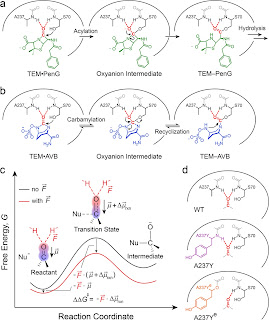
β-Lactamases Evolve against Antibiotics by Acquiring Large Active-Site Electric Fields

Zhe Ji and Steven G. Boxer

Journal of the American Chemical Society 2022 144 (48), 22289-22294

DOI: 10.1021/jacs.2c10791

A compound bound covalently to an enzyme active site can act either as a substrate if the covalent linkage is readily broken up by the enzyme or as an inhibitor if the bond dissociates slowly. We tracked the reactivity of such bonds associated with the rise of the resistance to penicillin G (PenG) in protein evolution from penicillin-binding proteins (PBPs) to TEM β-lactamases and with the development of avibactam (Avb) to overcome the resistance. We found that the ester linkage in PBP–PenG is resistant to hydrolysis mainly due to the small electric fields present in the protein active site. Conversely, the same linkage in the descendant TEM–PenG experiences large electric fields that stabilize the more charge-separated transition state and thus lower the free energy barrier to hydrolysis. Specifically, the electric fields were improved from −59 to −140 MV/cm in an ancient evolution dating back billions of years, contributing 5 orders of magnitude rate acceleration. This trend continues today in the nullification of newly developed antibiotic drugs. The fast linkage hydrolysis acquired from evolution is counteracted by the upgrade of PenG to Avb whose linkage escapes from the hydrolysis by returning to a low-field environment. Using the framework of electrostatic catalysis, the electric field, an observable from vibrational spectroscopy, provides a unifying physical metric to understand protein evolution and to guide the design of covalent drugs.


Protein Electric Fields Enable Faster and Longer-Lasting Covalent Inhibition of β-Lactamases

Zhe Ji, Jacek Kozuch, Irimpan I. Mathews, Christian S. Diercks, Yasmin Shamsudin, Mirjam A. Schulz, and Steven G. Boxer

Journal of the American Chemical Society 2022 144 (45), 20947-20954

DOI: 10.1021/jacs.2c10791

The widespread design of covalent drugs has focused on crafting reactive groups of proper electrophilicity and positioning toward targeted amino-acid nucleophiles. We found that environmental electric fields projected onto a reactive chemical bond, an overlooked design element, play essential roles in the covalent inhibition of TEM-1 β-lactamase by avibactam. Using the vibrational Stark effect, the magnitudes of the electric fields that are exerted by TEM active sites onto avibactam’s reactive C═O were measured and demonstrate an electrostatic gating effect that promotes bond formation yet relatively suppresses the reverse dissociation. These results suggest new principles of covalent drug design and off-target site prediction. Unlike shape and electrostatic complementary which address binding constants, electrostatic catalysis drives reaction rates, essential for covalent inhibition, and deepens our understanding of chemical reactivity, selectivity, and stability in complex systems.


Covalent inhibitors of the RAS binding domain of PI3Ka impair tumor growth driven by RAS and HER2

Joseph E Klebba, Nilotpal Roy, Steffen M Bernard, Stephanie Grabow, Melissa A. Hoffman, Hui Miao, Junko Tamiya, Jinwei Wang, Cynthia Berry, ...